
Form & Function Studios
Osteology study
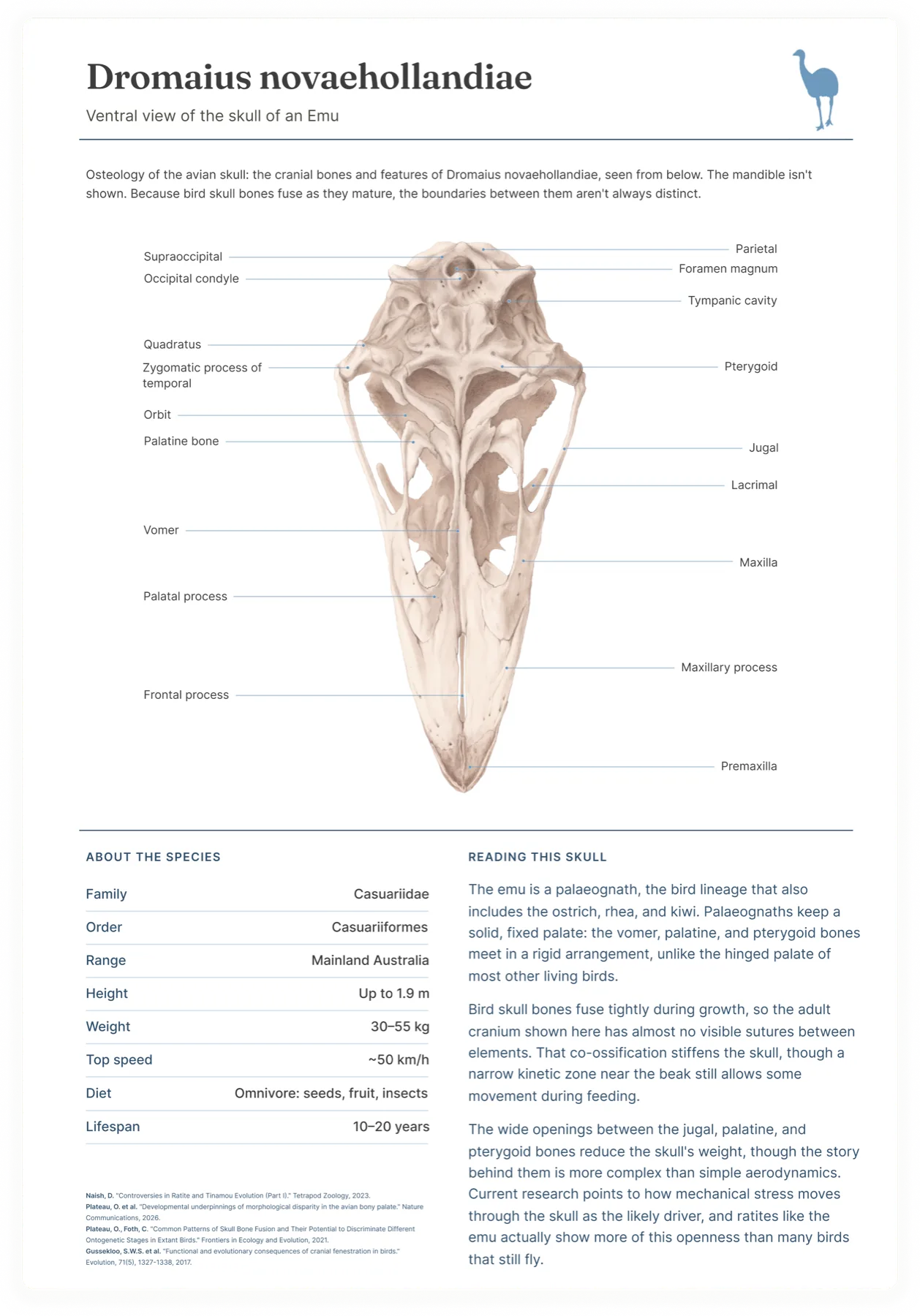
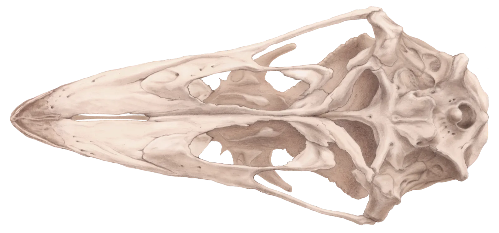
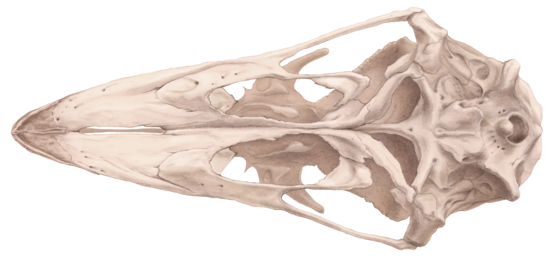
Most existing reference material shows the lateral or dorsal aspect of the skull, where the vomer, palatine and pterygoid are hidden behind the beak and braincase. It was therefore essential to draw from an actual specimen, which I was fortunate enough to have at hand.
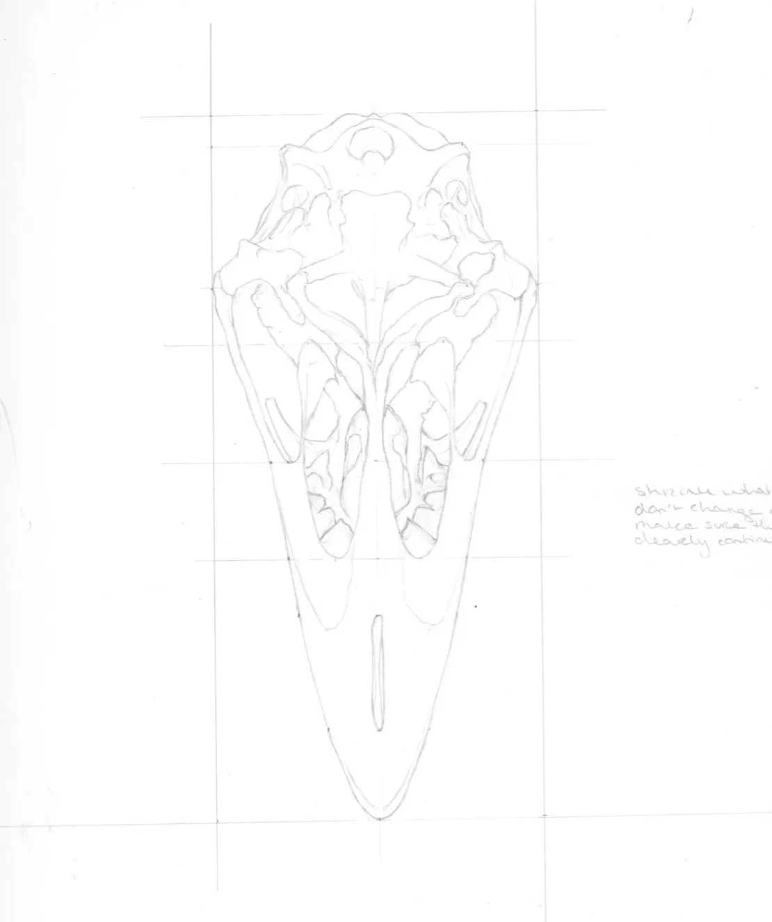
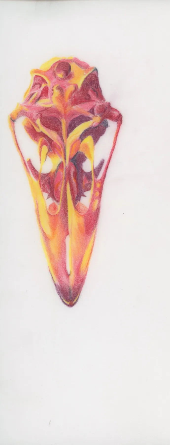
The skull was drawn 1:1, carefully measuring to get the proportions exactly right. After checking and double-checking all measurements, I created a value study. In this specific case it looks almost like a heat map, purely coincidental, based on the colours I chose that day. A value study is always good preparation before starting a final illustration or painting, but in this case I found it especially useful. The skull is such a delicate, light, and partially see-through structure that at first glance it's easy to think 'everything is light.' But there's always a range of values, and defining where the darkest darks and lightest lights fall, and everything in between, helps me capture the 3D shape when moving on to the final illustration.
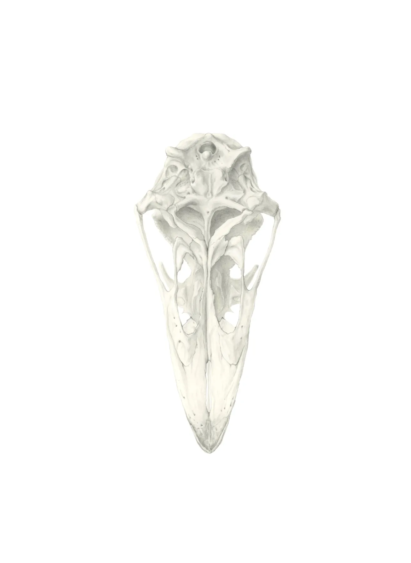
Just like the studies, the final skull was drawn in graphite at actual size. It was then coloured digitally by overlaying a solid 'skull' colour on top of the graphite pencil drawing, darkening some areas and highlighting others.