Form & Function Studios
Surgical study
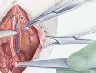
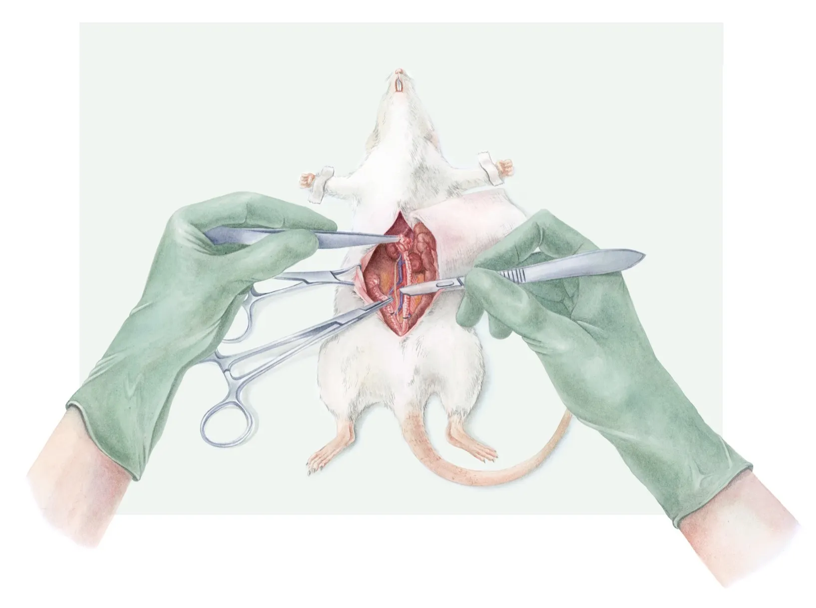
A procedure plate of an ovariohysterectomy in the rat, showing the abdominal approach: the midline incision, the reflected skin held with a hemostat, and the scalpel keeping tension along the cut margin.
There was no way to work from a single reference. The operating field is small, mostly obscured by the surgeon's own hands, and photographing over the shoulder gives you a viewpoint no reader needs. So the reference was assembled: a rat model to establish the animal's position and proportions, real surgical instruments to get the hands, the grip, and the metal right, and video footage of the actual procedure for what the tissue does when it is opened.
None of those three sources agreed on lighting, angle, or scale. Reconciling them is the actual work: a collage reference gives you accurate parts and no coherent whole, and the drawing has to invent the single consistent viewpoint that never existed.
Process
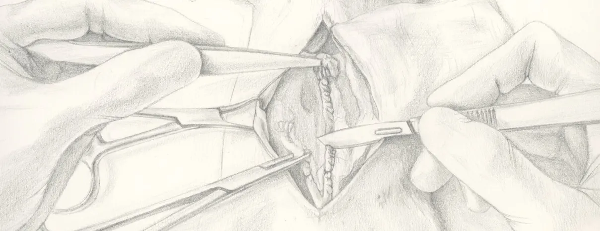
A graphite sketch came first, used as a composition sketch to puzzle together the parts assembled for the case study reference above. The main focus to make the whole thing come together was tissue behaviour, the light source, and reflection. The instrument I was using wasn't actually pulling the skin to the side, so this is something I had to fill in and create in a way that's believable. Imaginative realism, you could call it.
I settled on both the composition and the values in pencil before committing to any colours.
The finished illustration is made in watercolour, chosen because tissue is partly translucent and layered. It was then touched up digitally: edges cleaned, a few values pushed, the surgical steel sharpened, rather than repainted.