

Form & Function Studios
Case study
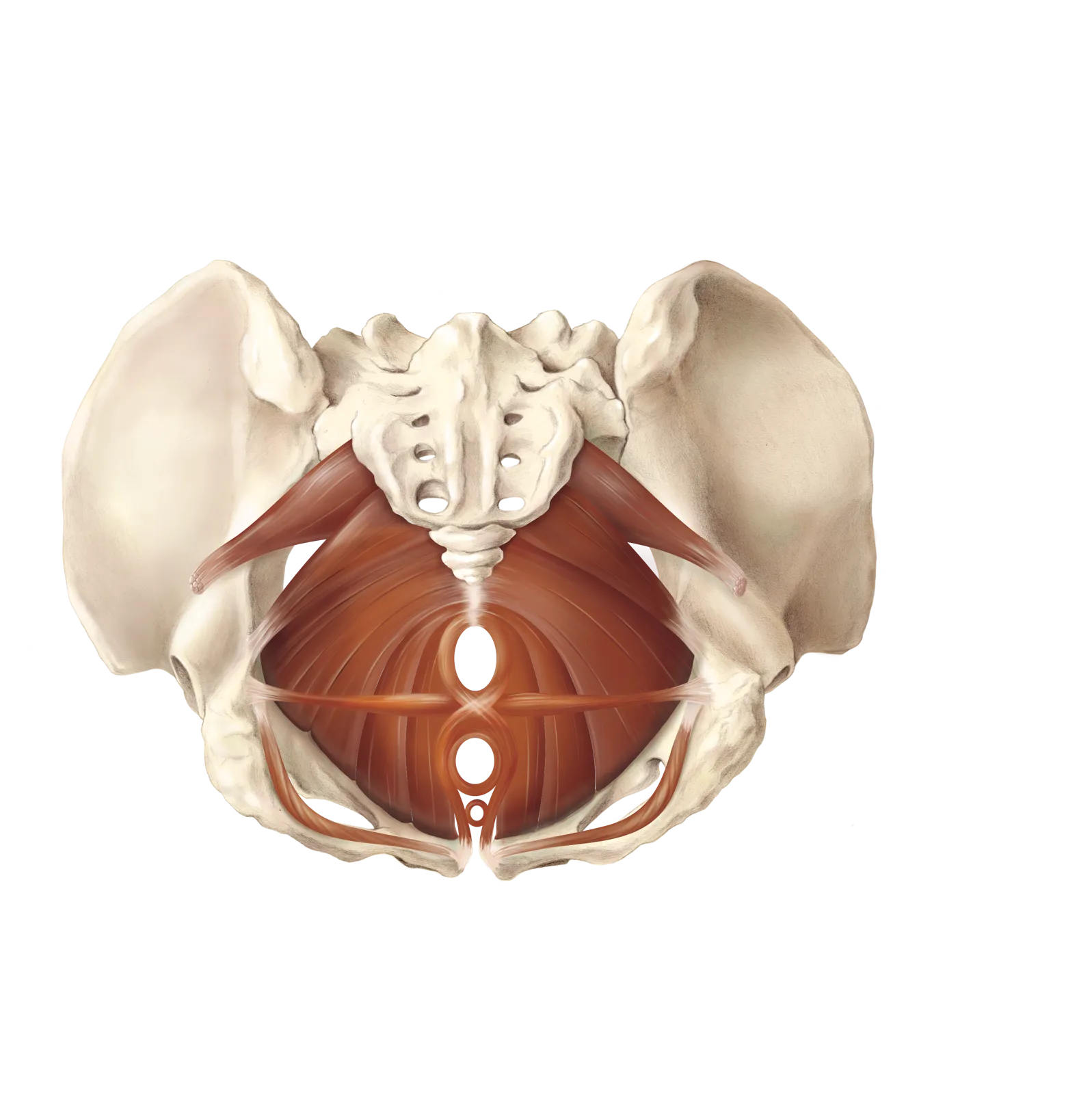
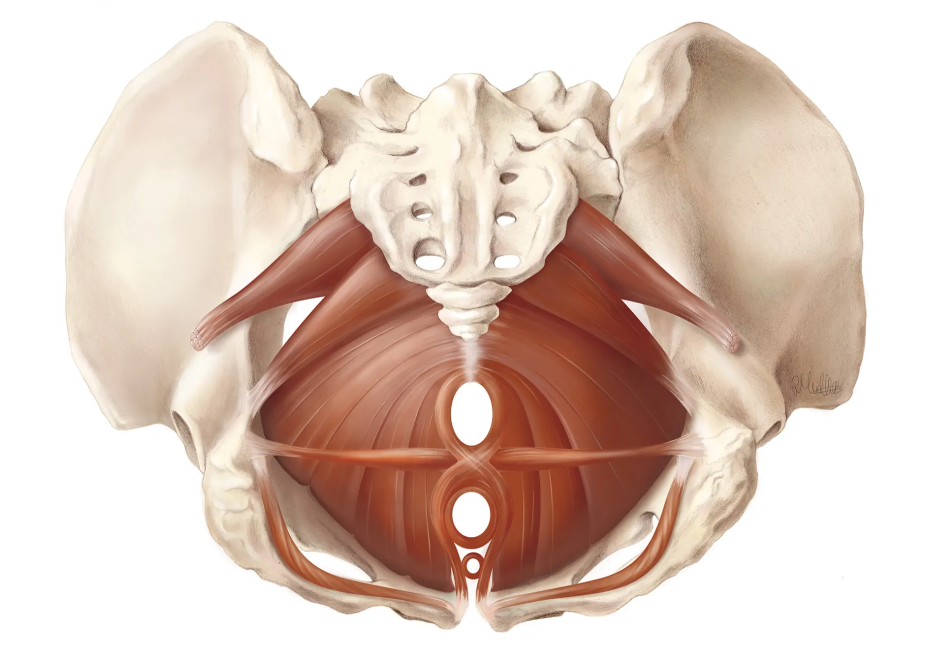
An illustration of the female pelvic floor made for Magee-Womens Research Institute in Pittsburgh, under gynaecological supervision, and drawn with access to a cadaveric dissection at the university research centre in Maastricht. The muscular layer is shown complete, in position, from below.
The pelvic floor is one of the hardest structures in the body to illustrate. It is a curved, three-dimensional sling of overlapping muscle that spans the pelvic outlet. No single viewpoint shows the whole of it and the layers don’t sit on top of one another as all illustrations will make you believe. The muscle-fibres are completely intertwined. In dissection the muscles are thin, pale, and fused at their borders. Textbook sources, as well as medical specialists contradict each other on the boundaries between individual muscles, and terminology varies between them.
The illustration therefore came out of three things running in parallel: the dissection in Maastricht, which gave the real proportions and the way the sheet attaches to the bony ring; extensive literature research, to resolve where the published sources disagreed; and repeated review with gynaecologists at Magee, who corrected the drawing against what they see in surgery. None of those alone would have been enough.
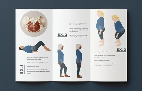
The pamphlet
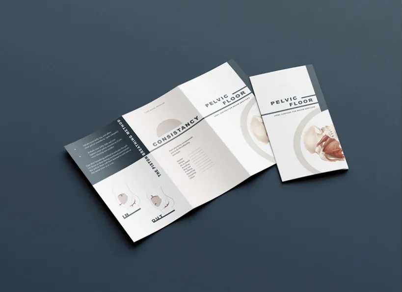
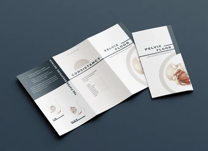
The plates were then set into a mock-up patient leaflet, to show the client how the same illustration could carry into patient education on pelvic floor training. Patients are routinely asked to exercise a group of muscles they have never seen, cannot easily feel, and are given no picture of. Showing them the actual structure changes the conversation: it makes the instruction concrete, and it makes patients feel taken seriously when they talk about the exercises they have been asked to do.
Another issue
When it comes to things like chronic pain, its about more than feeling understood, or pain relief, but giving patients the tools to manage their pain on the long term.
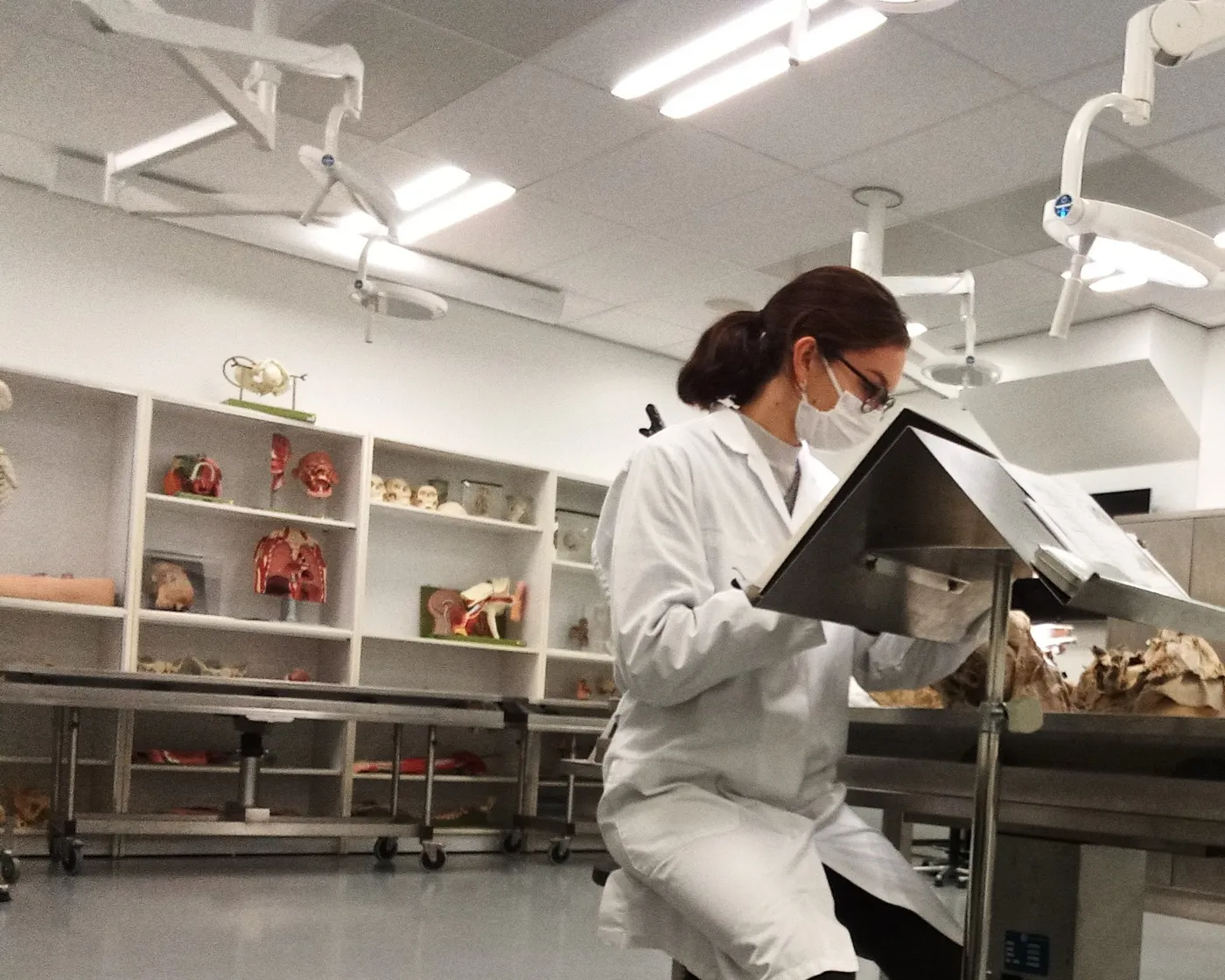
Working from a cadaveric dissection at the university research centre in Maastricht. These are the sketches made at the table: quick, unresolved.
Drawings made at a dissection are not preparatory studies for the final plate so much as understanding the complexity of the subject. The final illustration is a reconstruction, and these are in part what it was reconstructed from.