
Form & Function Studios

Case study
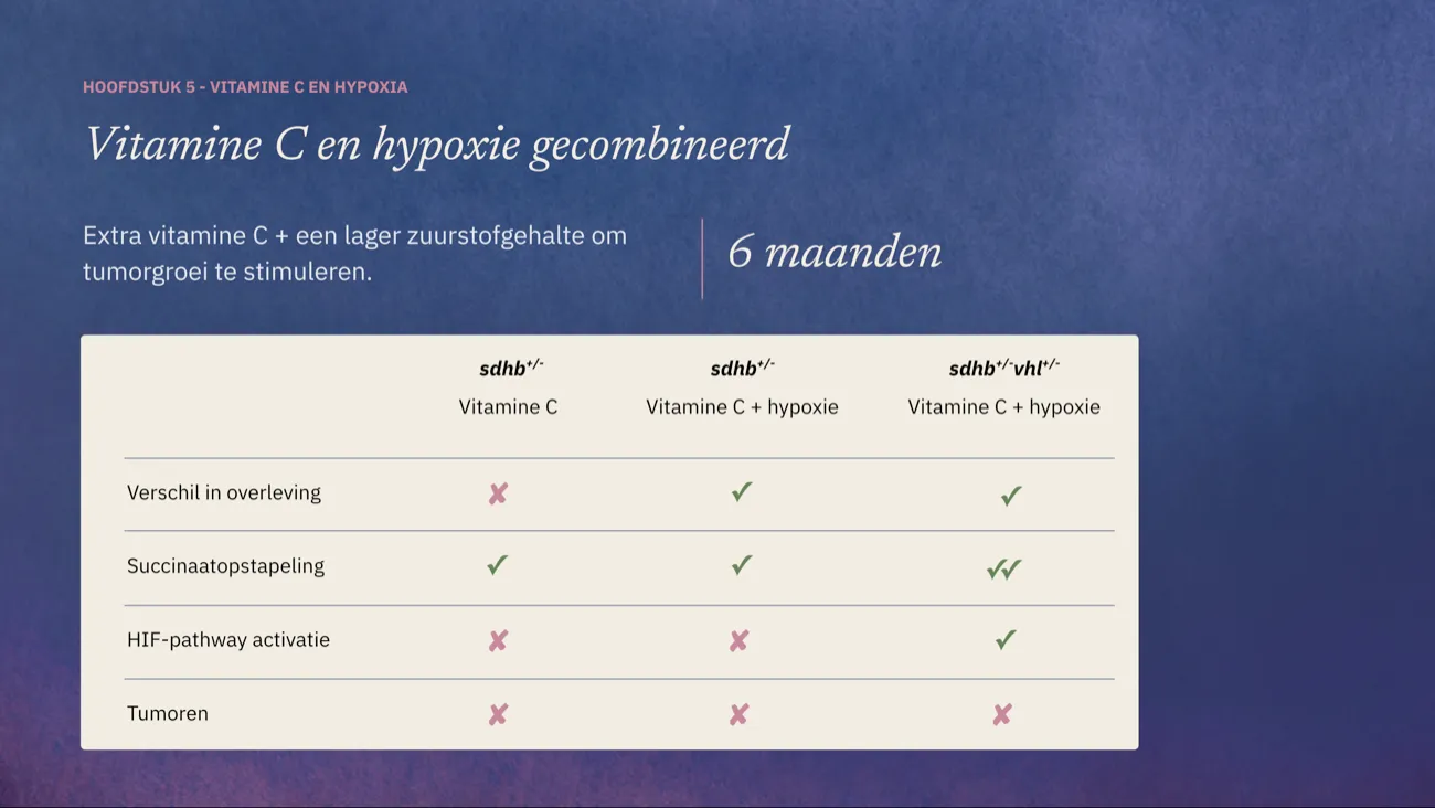
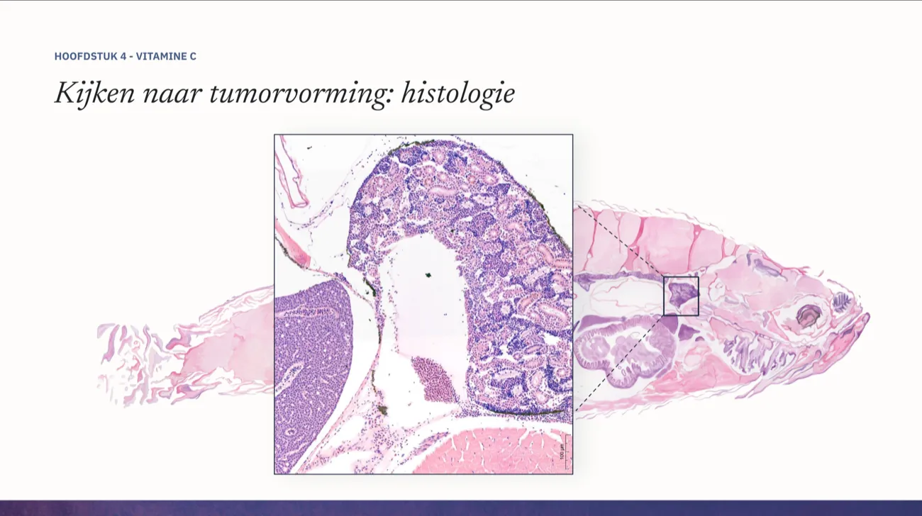
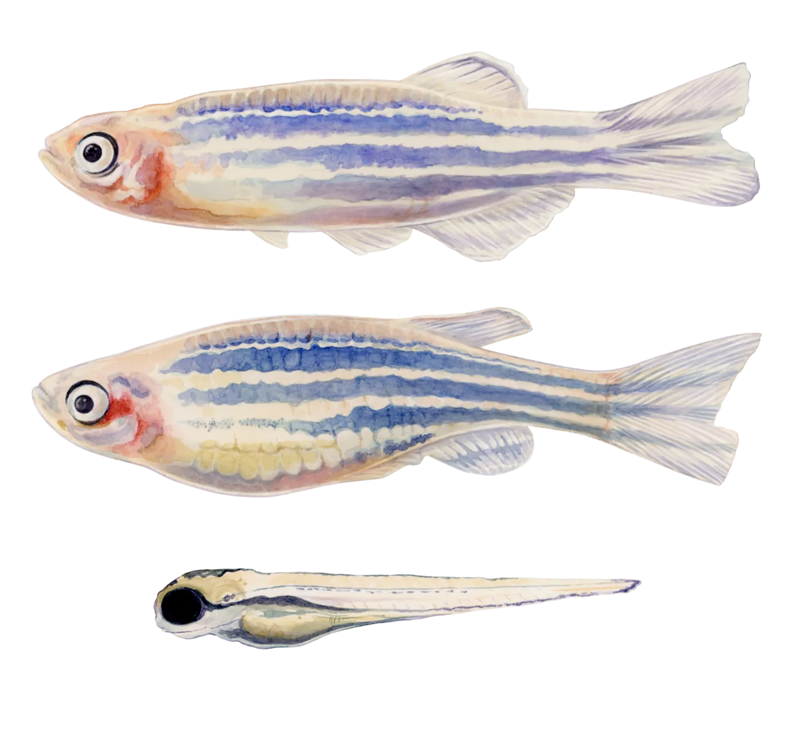
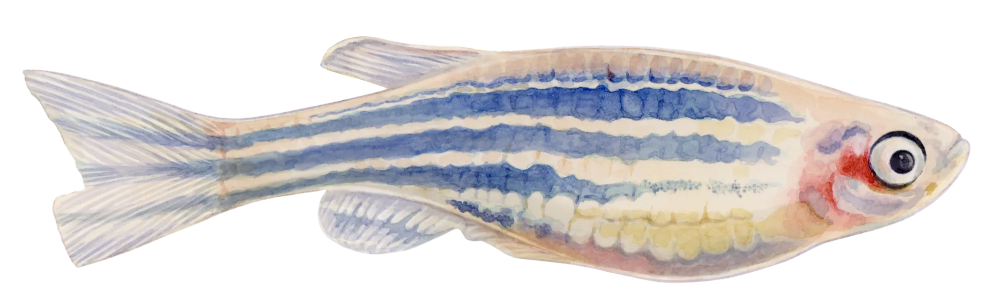
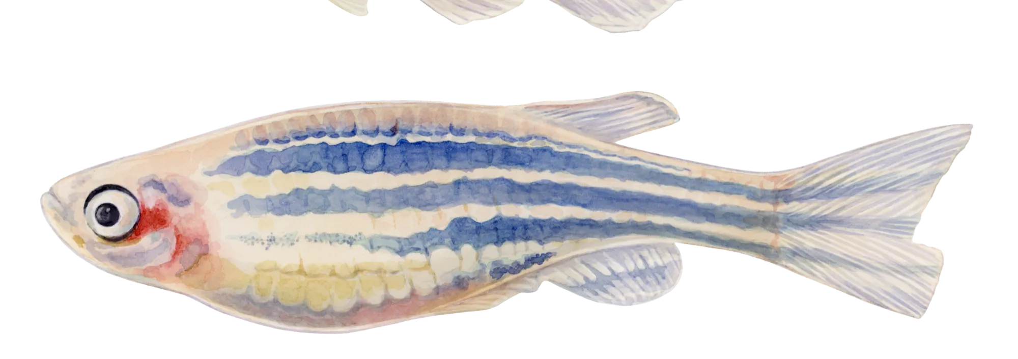
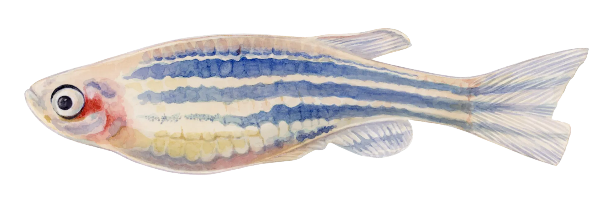
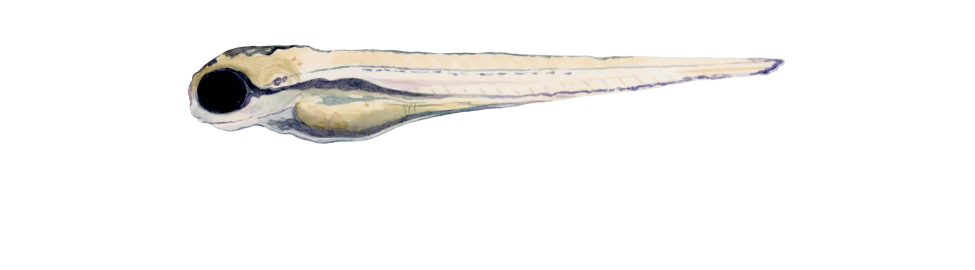
A set of developmental-stage illustrations for a PhD thesis on zebrafish embryogenesis, covering ten stages from cleavage through to free-swimming larva. The existing microscopy was accurate but hard to read at print size, and inconsistent between chapters — different angles, different magnifications, different labels for the same structures.
The brief wasn’t “make this pretty.” It was: make ten separate observations read as one continuous story.
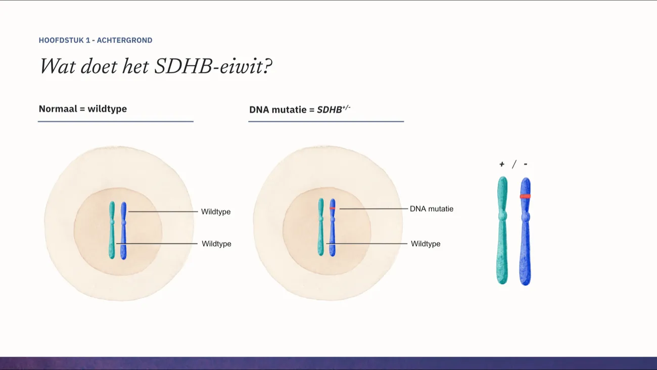
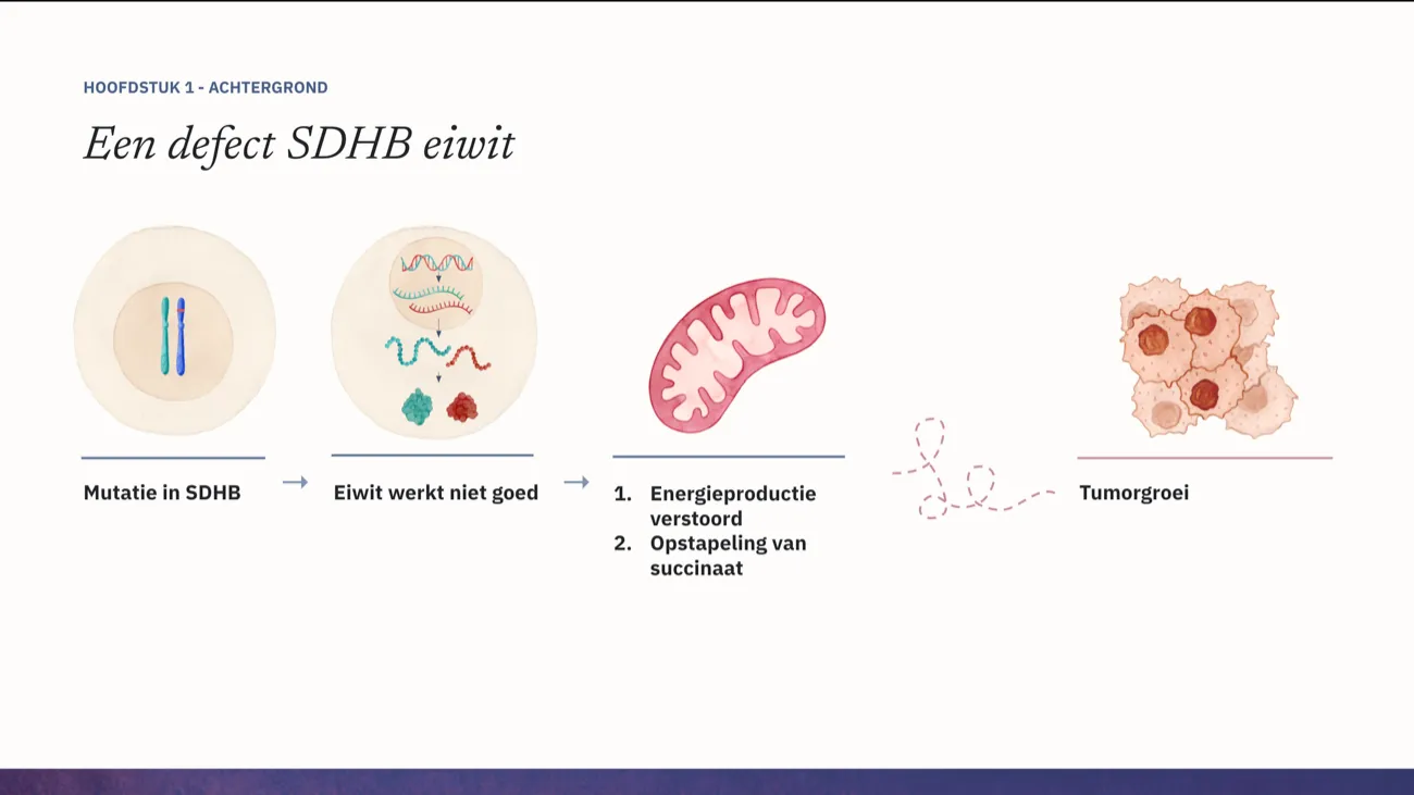
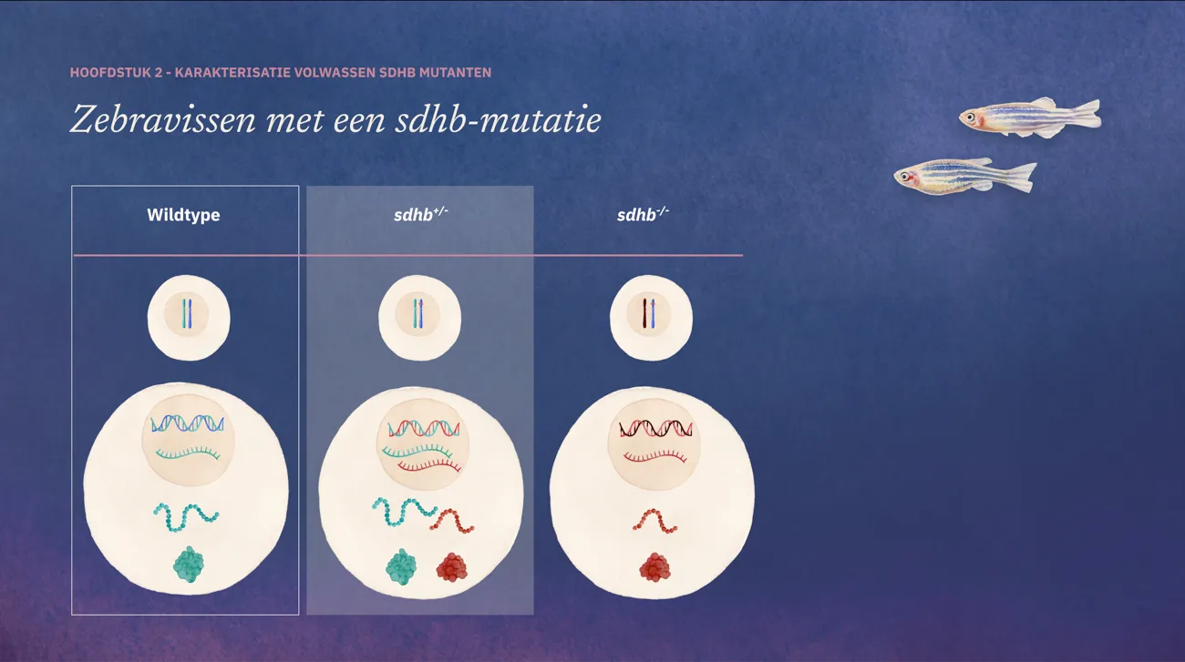
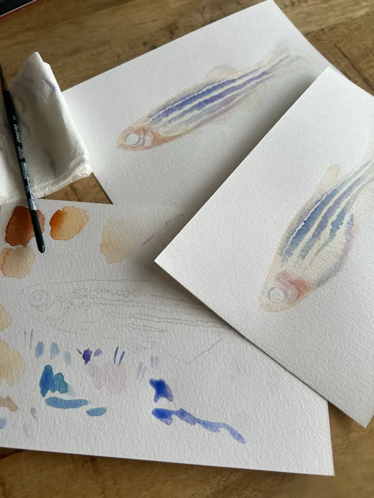
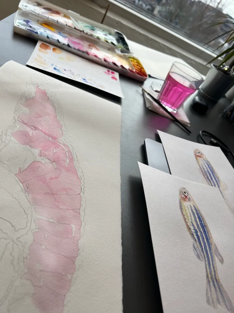
Each stage was redrawn from the original imaging with a shared palette, viewing angle, and labelling system. Structures that recur across stages keep the same colour throughout, so a reader can follow one organ forming across ten plates without re-learning the key each time.
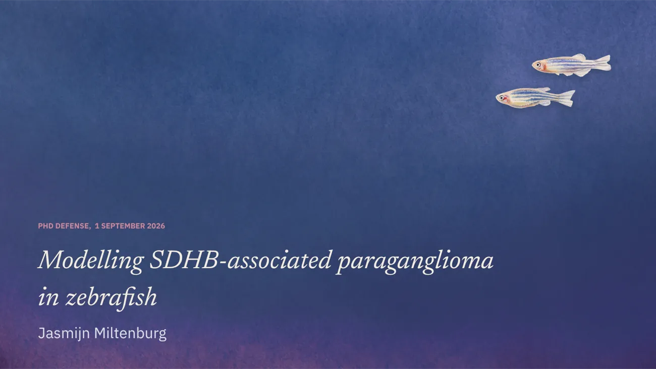
The series was then adapted three ways: full-bleed chapter openers for the printed thesis, simplified high-contrast versions for the defense slides, and CMYK-safe figures for the two papers those chapters became.
NoteFrom the supervisor
“The figures did more than illustrate the chapters — they made the argument legible. Three reviewers commented on them specifically.”